
Overview
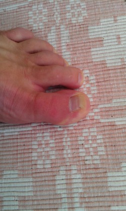
 Morton?s neuroma is a swollen nerve in the distal portion of the foot. The enlarged portion of the nerve represents scarring within the plantar nerve that occurs after chronic compression and/or repetitive injury. This may come about when the toes are squeezed together for too long, as can occur with the chronic use of high heels. The nerve that runs between your toes will swell and thicken. This can cause pain when walking. The symptoms of Morton?s neuroma can include burning pain in the foot, the feeling of a lump inside your foot, pain between the third and fourth toes typically but it can occur between other toes.
Causes
Some experts believe that other foot conditions may also be associated with Morton’s neuroma. This is because other conditions may cause the metatarsal bones to rub against the nerve in your foot. Foot problems that may increase your risk of developing Morton’s neuroma include abnormally positioned toes, high arches, where the arch or instep of your foot is raised more than normal, flat feet, low arches or no arches at all, bunions a bony swelling at the base of the toe. Hammer toe, where the toe is bent at the middle joint. Being active and playing sport can make the painful symptoms of Morton’s neuroma worse. In particular, running or sports that involve running, such as racquet sports, can place extra pressure on the nerve in your foot, which can aggravate the problem.
Symptoms
Symptoms associated with a neuroma include a dull burning sensation radiating towards the toes, a cramping feeling, or even a stinging, tingling sensation that can be described as being similar to an electric shock. It is often worse when wearing shoes with most people finding the pain disappears when removing their shoes.
Diagnosis
The physician will make the diagnosis of Morton’s neuroma based upon the patient’s symptoms as described above in an interview, or history, and a physical examination. The physical examination will reveal exceptional tenderness in the involved interspace when the nerve area is pressed on the bottom of the foot. As the interspace is palpated, and pressure is applied from the top to the bottom of the foot, a click can sometimes be felt which reproduces the patient’s pain. This is known as a Mulder’s sign. Because of inconsistent results, imaging studies such as MRI or ultrasound scanning are not useful diagnostic tools for Morton’s neuroma. Thus the physician must rely exclusively on the patient’s history and physical examination in order to make a diagnosis.
Non Surgical Treatment
There are various options for treating the condition, depending on its severity. Self-treatment. Here are some simple steps that may improve symptoms. Wear supportive shoes with a wide toe box. Do not lace the forefoot of the shoe too tightly. Shoes with shock-absorbent soles and proper insoles are recommended. Do not wear tight or pointed toed shoes or shoes with heels more than 2 inches high. Use over-the-counter shoe pads to relieve pressure. Apply an ice pack to the affected area to reduce pain and swelling. Rest your feet and massage the painful area. There are drugs that may temporarily relieve the pain and other symptoms of Morton?s neuroma. Long-term use of these medications is not recommended. Anti-inflammatory drugs-Nonsteroidal anti-inflammatory drugs, such as ibuprofen or aspirin, may be taken orally to reduce pain and inflammation. Anti-inflammatory drugs can also be administered by direct injection into the skin. Local anesthetic. An anesthetic injection will temporarily relieve pain by numbing the affected nerve. Orthotics. These are custom-designed shoe inserts that can reduce some of the pain associated with Morton?s neuroma. Sometimes padding is placed around the toe area, and tape is applied to hold the padding in place.
Surgical Treatment
If pain persists with conservative care, surgery may be an appropriate option. The common digitial nerve is cut and the Mortons neuroma removed. This will result is numbness along the inside of the toes affected, and there is a small chance the end of the nerve will form a Stump Neuroma. Approximately 75% of people receive symptom resolution for Mortons Neuroma with conservative care.


 Overview
 Symptoms
 Prevention
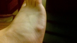 Overview
 Symptoms
 Prevention


